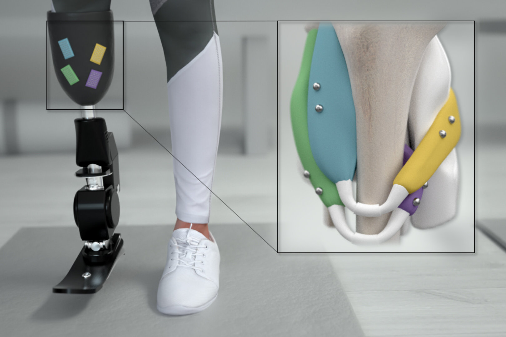
Today, prostheses and exoskeletons are controlled by electromyography. In other words, by recording the electrical activity during muscle contraction. It is neither intuitive nor human-like, it actually only shows the intention of the brain, not the reality of what the muscles are doing.
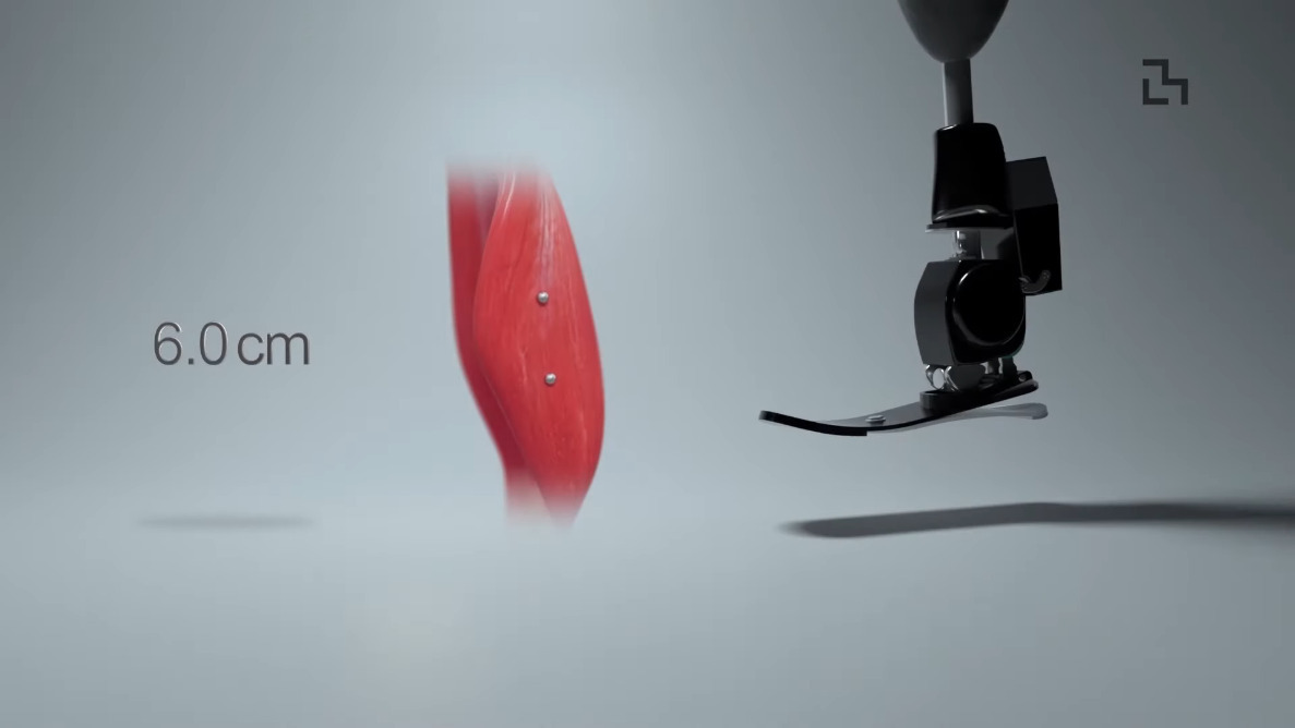
After embedding a pair of 3 mm diameter spherical magnets in a turkey calf, the researchers were able to detect muscle movement within 3 milliseconds with an accuracy of 37 microns, which is approximately the width of a human hair. They hope to try MM on humans in the next few years. If successful, this would be a good solution, because MM is cheaper, less invasive, and possibly permanent compared to the EMG method. Couple MM A new type of amputation surgery called AMI This provides a more comprehensive range of motion, reduces pain overall, and gives better control of the prosthesis. The future of prosthetics and rehabilitation looks very exciting. Be sure to check the video after the break.
There is more than one way to control a prosthesis, such as deep learning and somatosensory stimulation.
thanks for your reminder, [Qes]!
pass through Technological Exploration. Picture from MIT Media Lab.